


 Referrals & Patient Enquiries:


Twelve Lead ECG
Electrocardiography (ECG)
The electrocardiograph is a display of the heart’s electrical activation. It is often the starting point in the investigation of many cardiac conditions. It is a very routine non-invasive procedure and will usually be done each time you see a Cardiologist in the clinic. In addition to being a routine part of your physical examination and initial assessment the ECG is also used as a monitoring tool during many cardiac investigations and cardiac procedures.
How Is The Test Done?
There is no discomfort involved with having an ECG. You will be required to be bare chested and to lie flat on your back on an examination table. In some patients a small portion of chest hair will need to be shaved so the electrodes can be satisfactorily attached to the chest wall. The test will be usually performed by a technician or nurse but sometimes the Cardiologist may do the test themselves. Electrodes will be placed across your chest and attached to both wrists and both ankles. After these electrodes are attached you will lie quietly for a very short period while the ECG recording is taken.
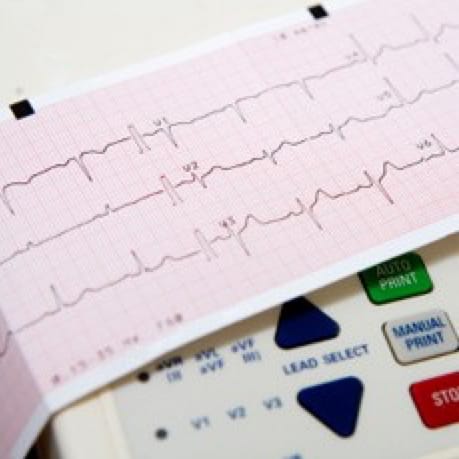
The result is a series of 12 wave forms recorded usually on ECG paper. These wave forms are produced by the electrical activity of your heart and supply a great deal of useful information.
The 12 lead ECG allows the Cardiologist to examine your electrical conduction system. It also allows us to assess the rhythm of the heart. In many diseased states the electrical signals of the heart are altered, and this can result in different variations of the electrical pattern of the heart and assist in diagnosis.